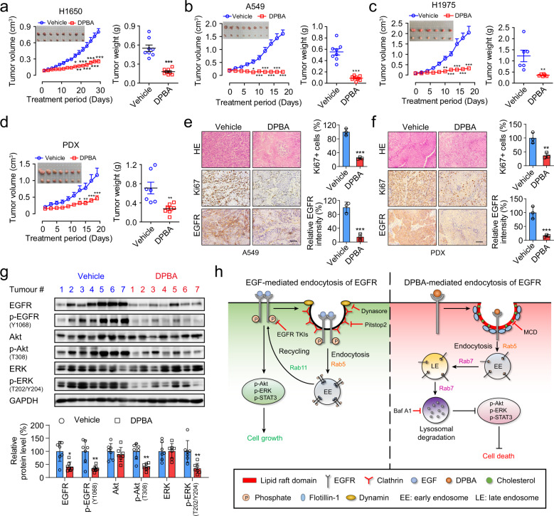
Fig. 5.
DPBA inhibits NSCLC growth in vivo. Tumour photos, tumour volume curves, tumour weights, and body weight curves for H1650 (a), A549 (b), H1975 (c), and primary lung PDX (d). *P < 0.05; **P < 0.01; ***P < 0.001 vs. the vehicle group. H&E staining and immunohistochemistry (IHC) staining for Ki67 and EGFR in A549 (e) and PDX (f) tumour tissues. Scale bar, 200 μm; **P < 0.01; ***P < 0.001 vs. the vehicle group, n = 3. g DPBA significantly suppressed EGFR pathway in PDX tumour tissues. *P < 0.05; **P < 0.01 vs. the vehicle group. h Schematic representation of the anticancer mechanism of EGFR small-molecule ligand DPBA

