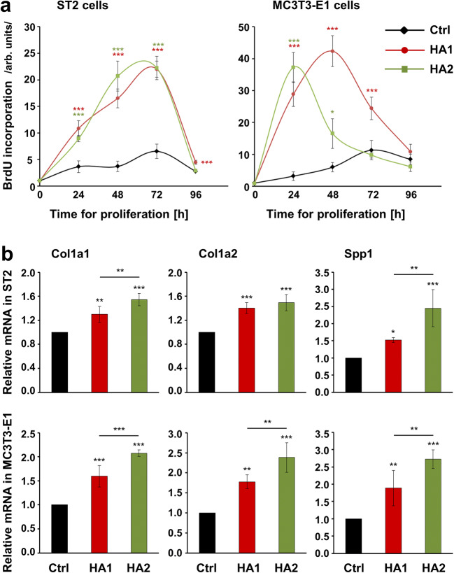
Fig. 1.
The two HA preparations strongly stimulate the growth of ST2 and MC3T3-E1 cells and enhance the expression of genes encoding bone matrix proteins. a Proliferation rates of HA-treated ST2 and MC3T3-E1 cells were assessed by BrdU incorporation into newly synthesized DNA immediately after plating (0 h) as well as at 24, 48, 72, and 96 h. Means ± SD from three independent experiments and significant differences to untreated control (Ctrl) cells at the time point 0; ***P < 0.001 and *P < 0.05 are shown. b Effect of HA1 and HA2 on Col1a1, Col1a2, and Spp1 mRNA levels in ST2 and MC3T3-E1 cells. Cells were treated with each of the two HA preparations for 24 h before total RNA was extracted and analyzed by qRT-PCR. Values normalized to Gapdh are expressed relative to the values of control (Ctrl) cells. Data represent means ± SD from three independent experiments. Significant differences to the respective control unless otherwise indicated, ***P < 0.001, **P < 0.01, *P < 0.05

