Fig. 3.
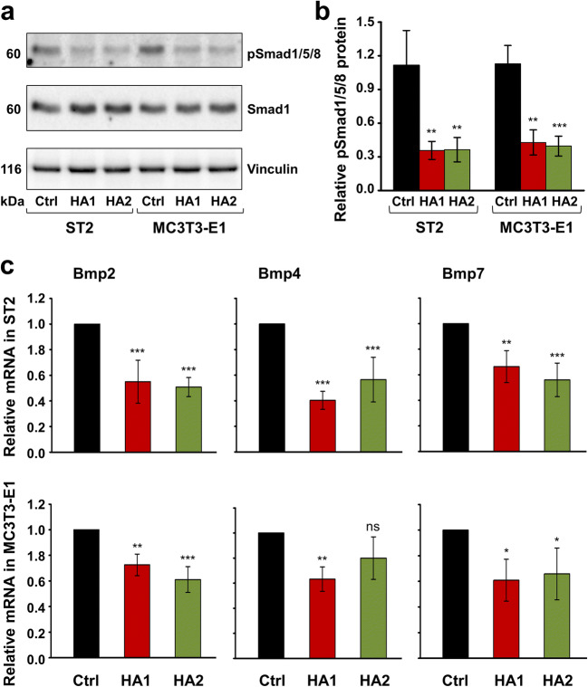
The two HA preparations inhibit BMP-activated Smad signaling in ST2 and MC3T3-E1 cells. a, b Immunoblot analysis of phospho-Smad1/5/8 (pSmad1/5/8) protein (a) in whole-cell extracts from ST2 and MC3T3-E1 cells treated with each of the two HA preparations. Blots for total Smad1 protein as well as the vinculin loading control are also shown a. The bar chart (b) represents densitometric quantification of the immunoblots. pSmad1/5/8 levels are normalized to the total Smad1 protein used as internal control. Data represent means ± SD from three independent experiments. Significant differences to the respective control (Ctrl) cells of each of the two cell lines, ***P < 0.001, **P < 0.01. c Effect of HA1 and HA2 on Bmp2, Bmp4, and Bmp7 mRNA levels in ST2 and MC3T3-E1 cells. Cells were treated with each of the two HA preparations for 24 h before total RNA was extracted and analyzed by qRT-PCR. Values normalized to Gapdh are expressed relative to the values of control (Ctrl) cells. Data represent means ± SD from three independent experiments. Significant differences to the respective control, ***P < 0.001, **P < 0.01, *P < 0.05, ns = not significant

