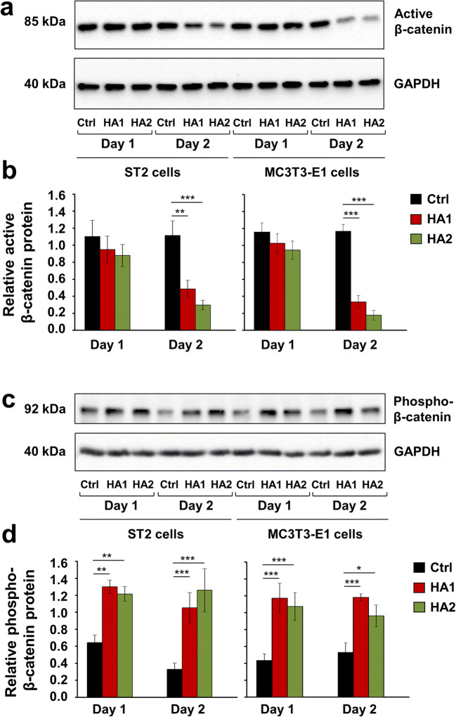
Fig. 7.
The two HA preparations inhibit Wnt signaling in ST2 and MC3T3-E1 cells. a–d Immunoblots of active β-catenin (a, b) and phospho-β-catenin (c, d) proteins in whole-cell extracts of ST2 and MC3T3-E1 cells treated with each of the two HA preparations. Cell lysates were collected on two consecutive days after the treatment. Anti-GAPDH served as loading control. Densitometric analyses (b, d) of the immunoblots shown in a and c. Active β-catenin (a, b) and phospho-β-catenin (c, d) protein levels are normalized to the GAPDH loading controls. Means ± SD from three independent experiments and significant differences to the respective control (Ctrl) cells, ***P < 0.001, **P < 0.01, *P < 0.05 are shown. No statistically significant differences between identically treated cells on days 1 and 2 after the stimulation were detected

