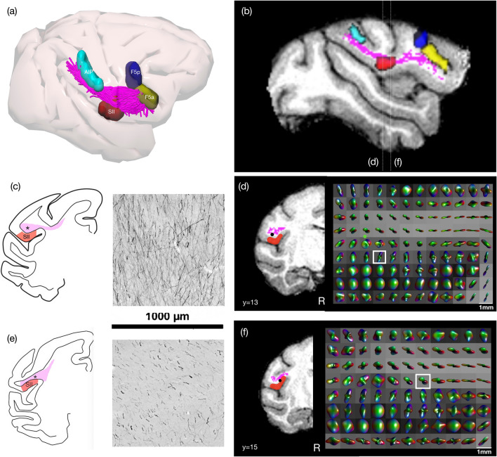
Fig. 6.
Comparison of labelled axons extending from the intraparietal area AIP in a macaque from a previous study (Case 30, Borra et al. 2008), with tractography and fibre ODF from spherical deconvolution modelling in one macaque from the Mount Sinai dataset. a Tractography of parieto-frontal connections of the LGNet running from AIP shown in 3D with respective sectors and b a sagittal slice showing this as a section. Comparison of c orderly axons within AIP-frontal projections running into SII which is also possibly reflected in a similar slice taken from the diffusion MR showing d fibre ODFs. e Orderly axons running anterior–posterior in a comparable section running within the core of the AIP-frontal projections and f fibre ODFs in a comparable coronal slice using diffusion MR. (N.B. in e there is also some cortical labelling)

