Figure 5.
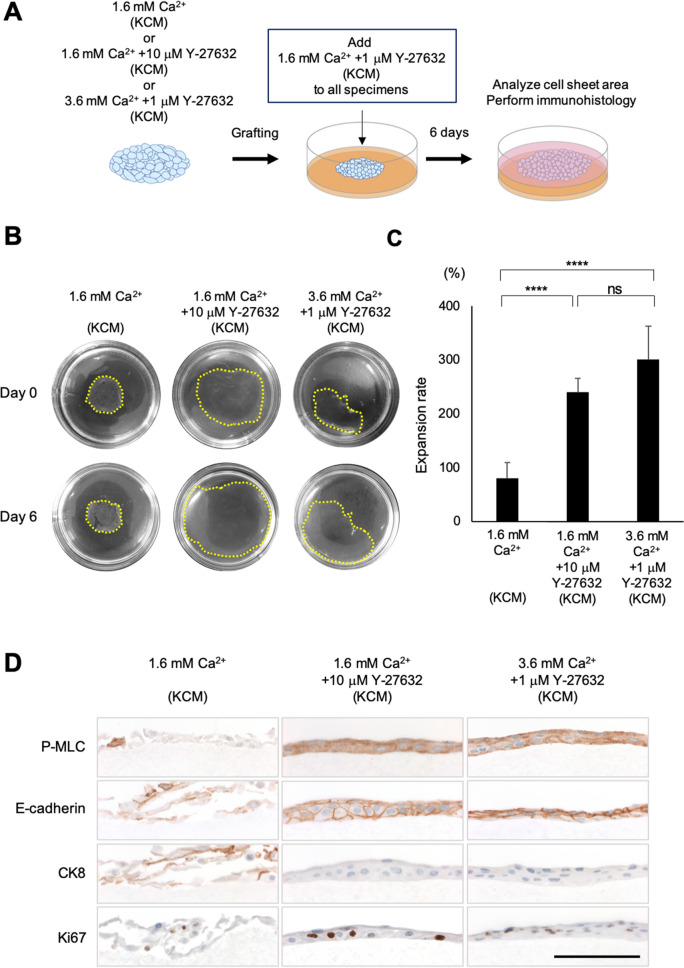
In vitro evaluation of the wound healing potential of cell sheets cultured in KCM containing 1.6 mM Ca2+, KCM containing 1.6 mM Ca2+ + 10 μM Y-27632, or KCM containing 3.6 mM Ca2+ + 1 μM Y-27632. (A) Schematic diagram of the in vitro cell sheet grafting assay. A harvested cell sheet was grafted onto a type I collagen gel and then cultivated for 6 days in KCM containing 1.6 mM Ca2+ and 1 μM Y-27632. (B) Representative images showing cell sheets before and 6 days after grafting onto type I collagen gels in 60-mm dishes. Yellow dotted line indicates the edge of each cell sheet. (C) Expansion rate 6 days after grafting onto the collagen gel. ****P < 0.0001; ns, not significant. (D) Immunohistological evaluation of the cell sheets at 6 days after grafting. Scale bar = 100 μm.

