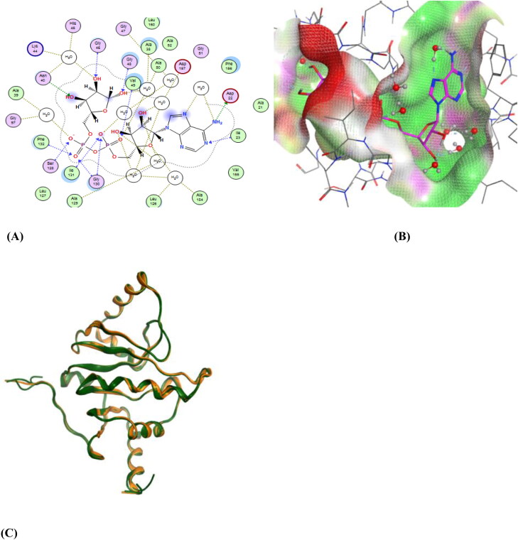
Figure 2.
(A) A 2D representation of the ligand-binding pocket showing attractive interactions between APR and amino acid residues. (B) A 3D representation of ligand APR (pink colour) interactions with amino acid residues with a surface representation. (C) Superposition of 6YWL and 6YWK showing structural similarity (Green colour – 6YWL, Orange colour – 6YWK).

