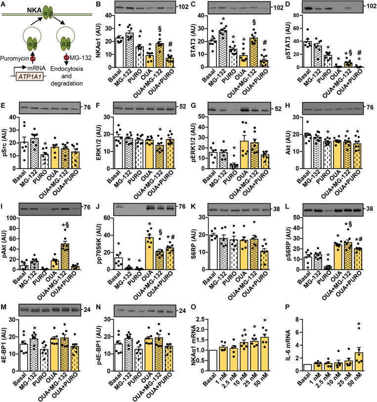
FIGURE 5.
Suppressive effects of ouabain on the abundance of NKAα1 and STAT3 in cultured human myotubes are mimicked by puromycin and blocked by MG-132. (A) Puromycin and MG-132 inhibit mRNA translation and protein degradation, respectively. (B–N): Human myotubes were treated in serum-free Advanced MEM with or without 1 μM MG-132 or 0.5 μg/ml puromycin (PURO) and/or 50 nM ouabain (OUA). MG-132 and puromycin were added 1 h before 24-h incubation with OUA. (B) α1-subunit of Na,K-ATPase (NKAα1), (C) the total STAT3, (D) phospho-STAT3 (Tyr705), (E) phospho-Src (Tyr527), (F) the total ERK1/2, (G) phospho-ERK1/2 (Thr202/Tyr204), (H) the total Akt, (I) phospho-Akt (Ser473), (J) phospho-p70S6K (Thr389), (K) the total S6RP, (L) phospho-S6RP (Ser235/236), (M) the total 4E-BP1 and (N) phospho-4E-BP1 (Thr37/46) were analyzed by immunoblotting. (O,P) Human myotubes were treated in serum-free Advanced MEM with different concentrations of OUA for 24 h. Expression of (O) NKAα1 mRNA and (P) interleukin-6 (IL-6) mRNA was determined with quantitative PCR. NKAα1 mRNA and IL-6 mRNA were normalized to three endogenous controls (PPIA mRNA, ACTB mRNA, and 18S rRNA). All values were normalized to respective Basal. Results are means with SEM (n = 6–8). *p < 0.05 vs. Basal, §p < 0.05 vs. MG-132, #p < 0.05 vs. puromycin.

