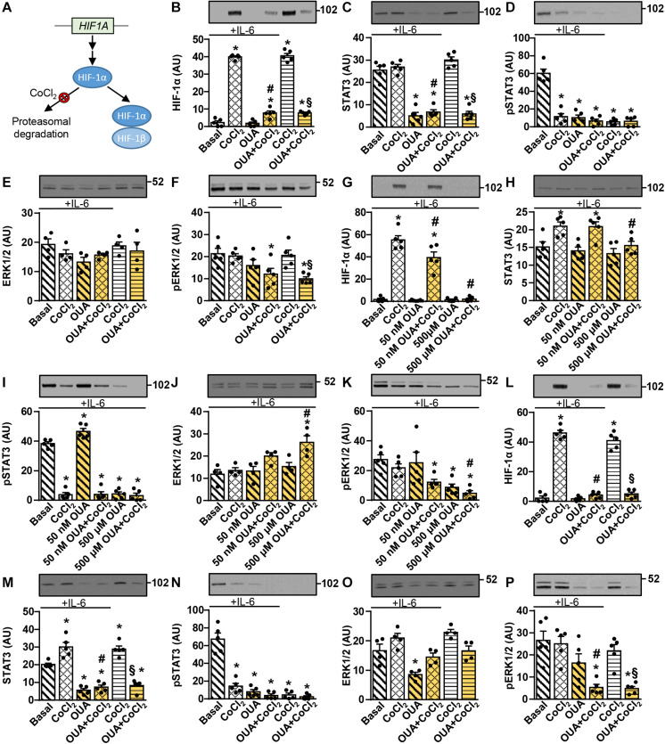
FIGURE 7.
Ouabain blocks CoCl2-induced upregulation of HIF-1α in cultured human myotubes and myoblasts. (A) Under normoxic conditions HIF-1α is continuously hydroxylated (not shown) and degraded in the proteasome. CoCl2 blocks HIF-1α hydroxylation, thus suppressing its proteasomal degradation, which leads to heterodimerization with HIF-1β. (B–F) Human myotubes were incubated in Advanced MEM with 2% FBS with or without 50 nM ouabain (OUA) for 20 h. During the next 4 h cells were incubated in serum-free Advanced MEM with or without 50 nM OUA and/or 250 μM CoCl2. During the final 15 min, cells were treated with or without 50 ng/ml interleukin-6 (IL-6). (G–K) Human myoblasts were incubated in serum-free Advanced MEM with or without 50 nM or 500 μM OUA and/or 250 μM CoCl2 for 4 h. Cells were treated with 50 ng/ml IL-6 during the final 15 min. (L–P) Human myoblasts were incubated in Advanced MEM with 10% FBS with or without 50 nM OUA for 20 h. During the next 4 h cells were incubated in serum-free Advanced MEM with or without 50 nM OUA and/or 250 μM CoCl2. During the final 15 min, cells were treated with or without 50 ng/ml IL-6. (B,G,L) HIF-1α, (C,H,M) the total STAT3, (D,I,N) phospho-STAT3 (Tyr705), (E,J,O) the total ERK1/2 and (F,K,P) phospho-ERK1/2 were measured by immunoblotting. Results are means with SEM (n = 4–5). *p < 0.05 vs. Basal; #p < 0.05 vs. CoCl2 + IL-6; §p < 0.05 vs. CoCl2.

