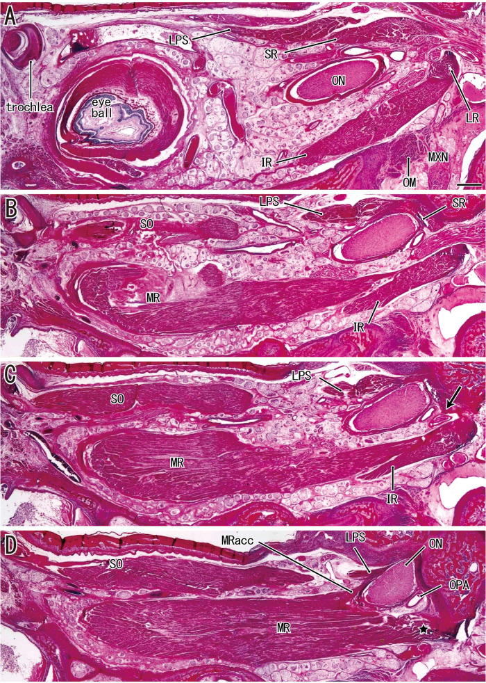
Figure 1.
Sagittal sections along the long axis of the orbit in a fetus with a CRL of 276 mm. (A) The most lateral site and (D) the most medial site. (A) The most posterior part of the LR. (B and C) The SR and LPS originate from the superior margin of the exit of the optic nerve (ON). (C, arrow) The inferomedial end of the SR origin. (D) The major and accessory heads of the MR (MRacc). All images were prepared at the same magnification (scale bar in A, 1 mm; ×1 objective). MXN, maxillary nerve; OM, orbital muscle (smooth muscle); OPA, ophthalmic artery.

