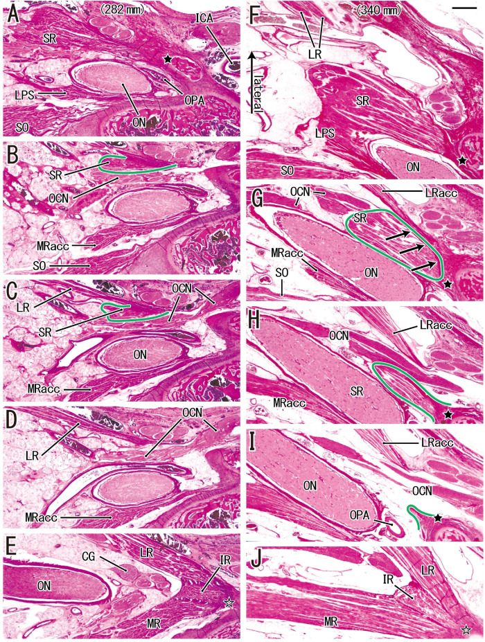
Figure 5.
An additional origin of the SR from a part of the LR in a fetus with a CRL of 282 mm (A–E) and a fetus with a CRL of 340 mm (F–J). (A and F) The most superior site of each fetus. (A, F, and I) The SR originates from the sphenoid (black star in each image). (E and J) The LR, IR, and MR have a common origin (open star in each image). (B and C) An inferolateral marginal part of the SR (surrounded by a green line) originates from the superior margin of the LR. Much inferiorly (1.0 mm), (E) shows that major parts of the LR and MR sandwich the IR origin. In contrast, (G) shows that an inferior part of the SR (surrounded by a green line) originates from an additional head of the LR (arrows). (H and I) The accessory head (LRacc) is detached from the bone. Much inferiorly (1.3 mm), (J) shows a major head of the LR. All images were prepared at the same magnification (scale bar in F, 1 mm; ×1 objective). ICA, internal carotid artery; MRacc, accessory head of the MR; OCN, oculomotor nerve; ON, optic nerve; OPA, ophthalmic artery.

