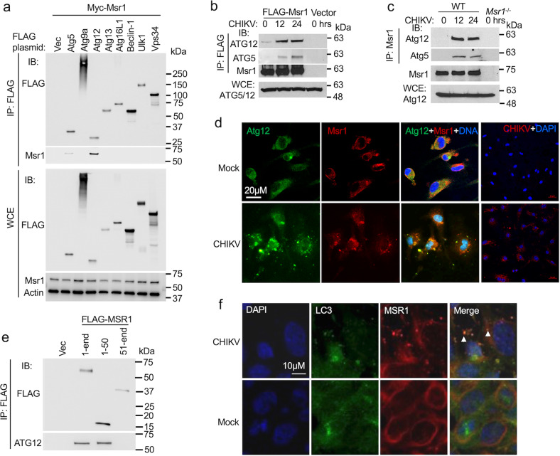
Fig. 3. CHIKV infection induces MSR1 interaction with the ATG5-ATG12 complex.
a Co-immunoprecipitation (co-IP) of FLAG-tagged mouse autophagy proteins with Myc-Msr1 expressed in HEK293 cells using a mouse monoclonal anti-FLAG antibody, followed by immunoblotting (IB) with a mouse monoclonal anti-FLAG and rabbit anti-Msr1 antibody. b Co-IP of FLAG-Msr1 (mouse) with endogenous human ATG5-ATG12 complex expressed in HEK293. CHIKV infection: MOI of 1. WCE: whole-cell extract. c Co-IP of endogenous Msr1 with the Atg5-Atg12 complex from bone marrow-derived macrophages (BMDMs) following CHIKV infection at a MOI of 10 for 12 and 24 h. The IP was carried out with a rabbit anti-Msr1 antibody cross-linked to protein A/G agarose beads. Msr1−/− cell serves as a negative control. d Immunofluorescence staining for Atg12 and Msr1 in BMDMs infected without (mock) /with CHIKV at a MOI of 10 for 12 h. Atg12 and Msr1 were stained by a mouse anti-Atg12 and rabbit anti-Msr1, followed by an Alexa Fluor-488 (green) and -594 (red)-conjugated secondary antibody respectively. The cells were counterstained for nuclei by DAPI (blue). The yellow punctae in the overlay indicate co-localizations. Magnification: ×400. e Co-IP of FLAG-MSR1 (human) or its fragment (aa1-50, 51-end) with endogenous ATG5-ATG12 complex expressed in HEK293 cells. CHIKV infection: MOI of 1 for 12 h. f Microscopic images of immunofluorescence staining for LC3B and MSR1 in trophoblasts without (mock)/infected with CHIKV at a MOI of 0.5 for 12 h. The yellow punctae in the overlay indicate co-localizations. Magnification: ×400. The uncropped immunoblots for all figures can be found in Supplementary Fig. 3.

