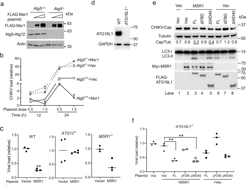
Fig. 4. Inhibition of CHIKV replication by MSR1 is dependent on ATG5-ATG12-ATG16L1.
a, b Repression of CHIKV replication by MSR1 requires ATG5. Mouse primary embryonic fibroblasts were transfected with either 0.2 or 1.5 µg of empty vector (Vec) or FLAG-Msr1 (mouse) expression plasmid. Twenty-four hours later the cells were then infected with CHIKV (multiplicity of infection MOI = 0.5). a Immunoblots showing FLAG-Msr1 (mouse) and Atg5-Atg12 expression at 24 h after plasmid DNA transfection. b Quantitative RT-PCR analyses of intracellular CHIKV RNA. N = 2. c Repression of CHIKV replication by MSR1 requires ATG12. Human trophoblasts were transfected with either 0.2 µg of empty vector or a FLAG-MSR1 (human) expression plasmid. Twenty-four hours later, the cells were then infected with CHIKV MOI = 0.5 for 12 h. Intracellular CHIKV RNA was quantitated by RT-PCR. n = 3 biologically independent samples. d Generation of human ATG16L1−/− trophoblasts by CRISPR-Cas9. The immunoblot shows ATG16L1 knockout efficiency. e Repression of CHIKV replication by MSR1 requires the FBD domain of ATG16L1. ATG16L1−/− trophoblasts were transfected with the indicated combinations of expression plasmids (human gene) and vector (Vec) (50 ng each) respectively. After 24 h, the cells were infected with CHIKV at a MOI of 0.5 for 16 h. FL: full-length, ΔFBD: FBD domain deletion, ΔWD40: WD40 domain deletion of ATG16L1. The immunoblots show CHIKV Capsid and cellular protein expression. n = 3 biologically independent samples. The small horizontal line: the median of the result. *p < 0.05, **p < 0.01 (Student’s t-test). The uncropped immunoblots for all figures can be found in Supplementary Fig. 3.

