
Figure 1. Correlation of radical prostatectomy histopathology with mpMRI.
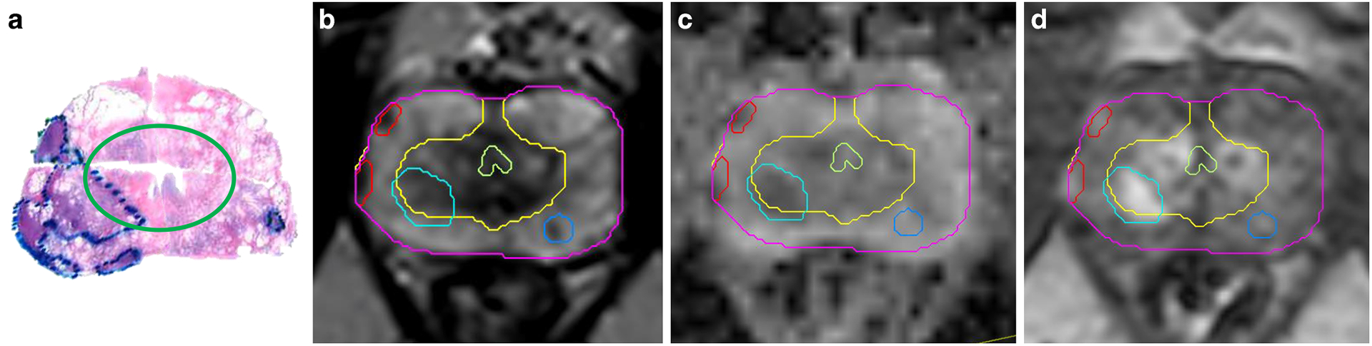
a) Transverse pseudo whole mount section of H&E-stained radical prostatectomy from a patient with prostate cancer. The areas of the tumors are marked by pathologist. The green ellipse separates the peripheral (outer) and transition zones of the prostate; b) Corresponding T2-weighted axial MRI image is shown. The prostate is contoured in magenta. The two zones of the prostate have different intensities: the T2 signal is bright on the peripheral zone (yellow contour), due to the fact that the peripheral zone contains mainly prostatic acini and ducts and the prostatic fluid in their lumina rises a strong signal. Alternatively, the transition zone has a higher proportion of prostatic stroma. The light green structure is the urethra. Correspondingly, on the histopathology sample, the transition zone is surrounded by a porous tissue related to the frequent glandular luminae in the peripheral zone. The tumors (red and shades of blue) on T2-weighted MRI are hypo-intense. c) Apparent Diffusion Coefficient (ADC) image. ADC is calculated from Diffusion weighted imaging (DWI) - a specialized acquisition technique which highlights areas with restricted microscopic motion of water molecules. Tumors on ADC are also hypo-intense. d) The early enhancing image in the Dynamic Contrast Enhanced (DCE-) MRI. DCE-MRI highlights the tumor vasculature and areas of the tumor are of higher intensity.