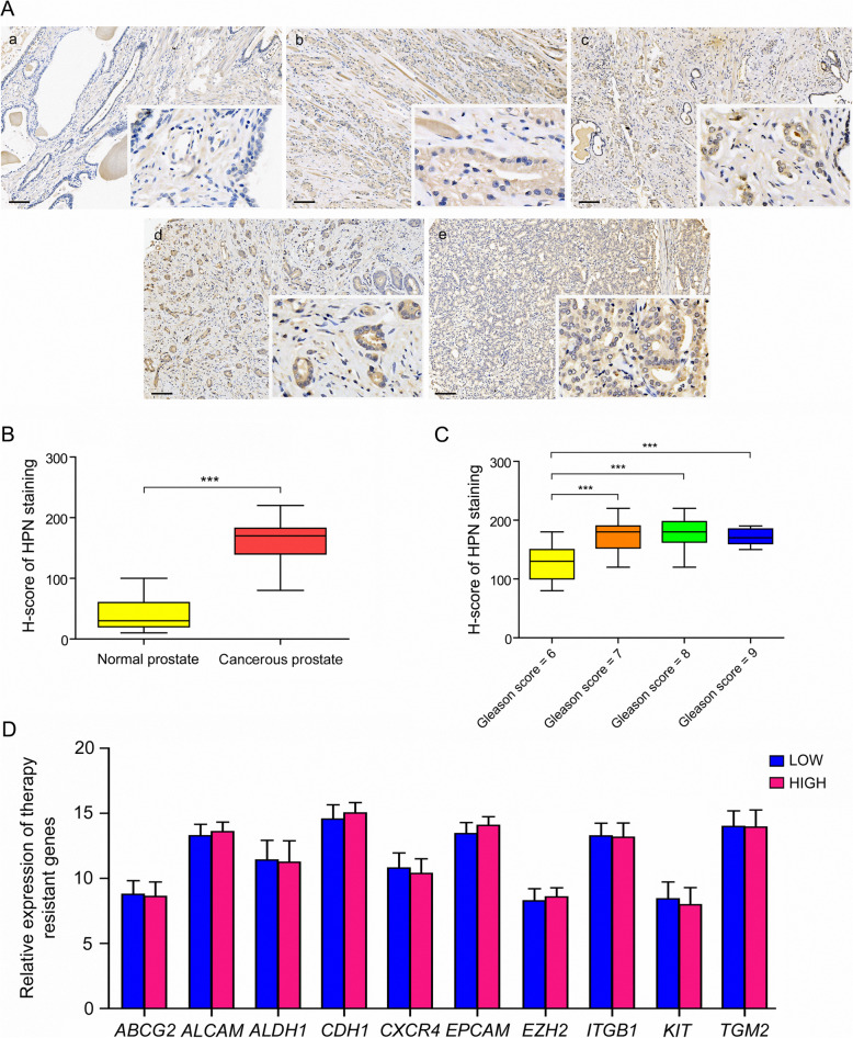
Fig. 7.
Validation of HPN expression in PCa tissue array. a Immunostaining of HPN in normal prostate and cancerous prostate with different pathology grading. Positive signals with anti-HPN were stained in brown. Cell nucleus were stained with hematoxylin and presented blue in PCa tissue sections. a normal prostate, b cancerous prostate with a Gleason score of 6, c cancerous prostate with a Gleason score of 7, d cancerous prostate with a Gleason score of 8, e cancerous prostate with a Gleason score of 9. b H-score of HPN staining in normal prostate and cancerous prostate. c H-score of HPN staining in PCa tissues with different pathology grading. d Relative expression of therapy-resistant markers in PCa patients with low and high expression levels of HPN, LOW patients with low expression of HPN; HIGH patients with low expression of HPN

