Figure 4.

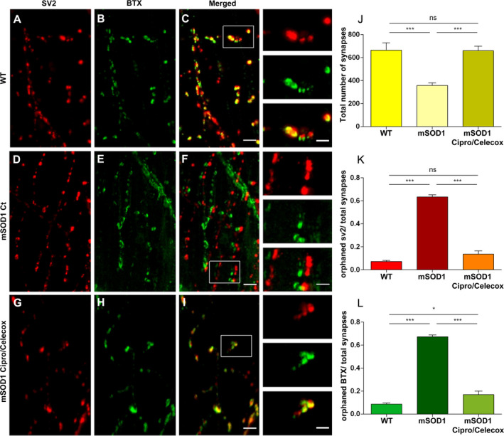
Cipro/Celecox rescued orphaned pre‐ and post‐synaptic components in mSOD1 larvae. (A‐I) Representative images of single ventral root projection double‐labeled for SV2 (presynaptic marker, A, D, G), BTX (postsynaptic marker, B, E, H), and colocalization of α‐SV2 and α‐BTX (merged, C, F, I), in WT (A–C), mSOD1 control (D‐F) and mSOD1 treated with 100 µmol/L Cipro/ 1 µmol/L Celecox (G‐I). (Scale bar = 30 µm, scale bar in insets = 15 µm). (J) Quantification of the number of synapses formed, quantified as the number of colocalized SV2 and BTX puncta. (K) Quantification of orphaned presynaptic SV2 puncta over total number of SV2 puncta. (L) Quantification of orphaned postsynaptic α‐BTX staining over total number of α‐BTX puncta. (ns = non significant; *P < 0.05; **P < 0.01; ***P < 0.001; one‐way ANOVA, Tukey post hoc test, n = 18 for WT and Cipro/Celecox treated mSOD1 fish and n = 36 for control mSOD1 fish).
