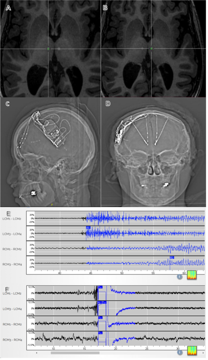
Figure 2.

(A) Merged MPRAGE‐CT neuronavigation targeting right CMT; (B) Merged MPRAGE‐CT neuronavigation targeting left CMT; (C) AP X‐ray of RNS in situ with frontal and centromedian thalamic depth electrodes; (D) Lateral X‐ray of RNS in situ, same patient; (E) Seizure detection on thalamic leads before stimulation is activated; (F) Ictal pattern detected, stimulated and stopped from development in thalamic leads. LCM, left centromedian, RCM, right centromedian. L,R CM1‐L,R CM2 = deeper electrodes; L,R CM3‐L,R CM4 = more superficial electrodes.
