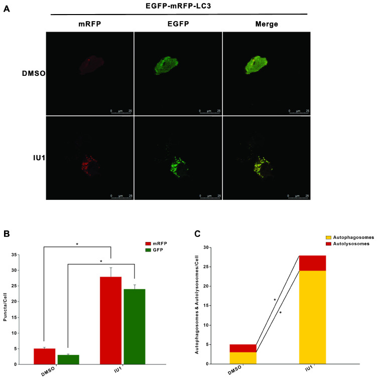
Figure 7.
The autophagy flux was induced when cells were treated with IU1. (A) The EGFP-mRFP-LC3 assays in vitro. HeLa cells were transfected with pBabe-EGFP-mRFP-LC3 vector for 48 h and were subjected to IU1 100 µM for 12 h. Representative images of fluorescent LC3 puncta are shown. (B) Mean number of GFP and mRFP dots per cell. (C) Mean number of autophagosomes and autolysosomes per cell. Results represent the means from at least three independent experiments. *p < 0.05; **p< 0.01. Scale bar represents 25 µm.

