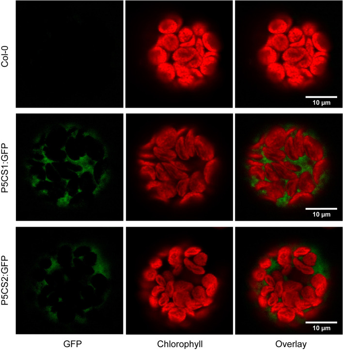
Figure 1.
Subcellular localization of P5CS1 and P5CS2. GFP and chlorophyll fluorescence images of mesophyll protoplasts isolated from wildtype and P5CS1:GFP or P5CS2:GFP expressing plants. Spectrally resolved confocal fluorescence images with 20 channels spanning 490 to 668 nm emission wavelength were used for spectral unmixing with GFP and chlorophyll reference spectra. The GFP images and the overlay demonstrate that fluorescence emission from chloroplasts was entirely attributable to chlorophyll, and GFP fluorescence was exclusively detected in the cytosol. Compare with the simulated channel splitting mode images in Supplementary Figure S1 .

