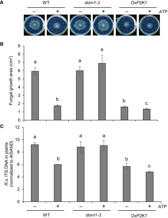
Figure 4.

Extracellular ATP reduces Rhizoctonia solani AG2-1 infection in roots. (A) Representative pictures of R. solani infection in MS plates with wild type (WT), dorn1-3, and OxP2K1 seedlings at 2 days post infection. (B) The area of fungal growth on the MS plates in (A). (C) The level of infection in the seedlings quantified by real-time PCR. Abundance is expressed as the ratios of the fungal-specific ITS region relative to the Arabidopsis reference gene AtSAND. The values are the means ± SE of three biological replicates. Different letters indicate significant differences at P < 0.05.
