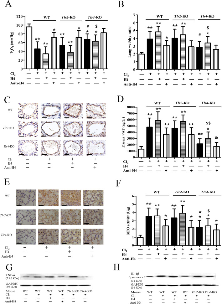
Fig. 3.
Roles of histone H4 and TLRs in Cl2 induced inflammatory lung injury. Twenty four hours after Cl2 exposure (400 ppm, 30 min) in WT, Tlr2-KO, and Tlr4-KO mice, PaO2 (3A) and lung wet/dry mass ratio (3B) were measured. Pulmonary endothelial activation was represented by P-selectin expression (3C) (Scale bars: 100 μm) and circulating vWF (3D). Pulmonary neutrophil infiltration was shown by Ly6G marker staining (3E) (Scale bars: 50 μm), and neutrophilic activation was indicated by MPO activity (3F). The protein levels of inflammatory cytokines TNF-α (3G) and IL-1β (3H) were measured by western blot. Histone H4 (10 mg/kg) or anti-H4 antibody (20 mg/kg) was delivered through the tail vein 1 h prior to Cl2 exposure. N = 6 independent replicates for all groups. *p < 0.05, **p < 0.01 compared with the control group; #p < 0.05, ##p < 0.01 compared with the WT mice treated solely with Cl2; $p < 0.05, $$p < 0.01 compared with the WT mice treated in the same manner (Cl2 + H4); &p < 0.05, &&p < 0.01 compared with the WT mice treated in the same manner (Cl2 + Anti-H4).

