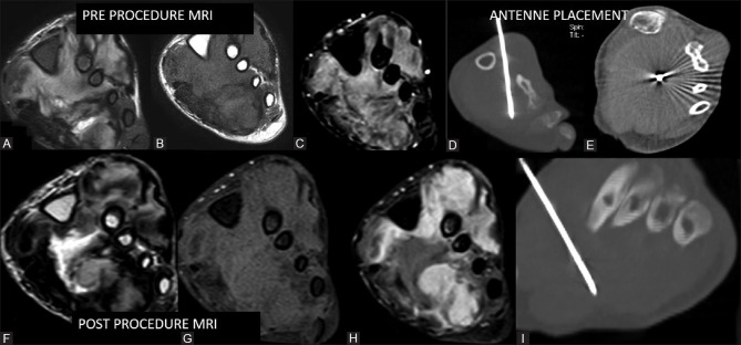
Figure 10 (A-I).
19-year-old male with history of surgery done twice and recurrence of swelling. Patient was started on metronomic chemotherapy but had progression of swelling and thus was referred for ablative therapy. Preprocedure MRI reveals soft tissue mass on the plantar aspect encasing metatarsal with iso- to hypo-intense on T2 (A) and shows heterogenous post-contrast enhancement (B and C). Antenna placement done (D, E and I). Small nonenhancing necrotic area is seen post microwave ablation (F-H)

