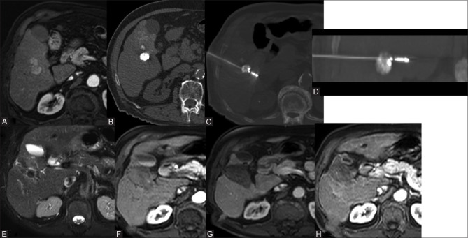
Figure 7 (A-H).
70-year-old male with multiple co-morbidities. CEMR reveals solitary lesion in the segment V/VI of liver showing arterial wash-in and venous wash out (A). Lipiodol TA TACE done, plain CT reveals good Lipiodol deposition (B), MW antenna was placed 1 cm beyond the lesion (C). Post MWA MR was done on follow-up (E-G) which reveals on T2 hypo intensity and no enhancement. 6-month follow-up also reveals no enhancement (H)

