

Abstract
Unilateral or bilateral agenesis of the parotid gland is an uncommon condition with unclear aetiology. Only 22 cases of unilateral salivary agenesis have been reported excluding the present case. We present a case of a 4-year-old female child who presented with complaints of slight discoloration of her tongue and was referred for MRI to rule out any vascular malformation. Imaging revealed a complete absence of the right parotid gland. Hypertrophy of the sublingual gland and hypoplasia of the parotid gland on the opposite side was also noted, an unusual finding as the contralateral parotid showed compensatory hypertrophy in the other reported cases.
Keywords: Bifid tongue, parotid, salivary agenesis, supernumerary teeth
Case Summary
A 4-year-old girl with complaints of slight discoloration of her tongue was referred for MR imaging to rule out any vascular malformation. The patient was otherwise asymptomatic. On physical examination, the presence of bifid tongue, supernumerary teeth on the alveolar border of the maxilla, a solitary mandibular incisor and a high-arched palate were noted. No other orofacial or digital defects were noted.
Imaging Findings
MRI with contrast enhancement was performed on a 3.0T MRI system. The study revealed a complete absence of the right parotid gland [Figure 1]. The left parotid gland was seen at its normal position but was reduced in size. The presence of bilateral accessory parotid tissue was also noted [Figure 2]. A well-defined soft tissue mass of 15 × 8 mm was seen in the right sublingual region, isointense to salivary gland tissue on all sequences, representing hypertrophied sublingual gland [Figure 3]. Bilateral submandibular glands appeared normal. No vascular abnormalities were noted.
Figure 1.
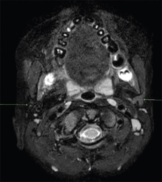
Axial T2 FATSAT image showing absent right parotid gland and small (hypoplastic) left parotid gland
Figure 2.
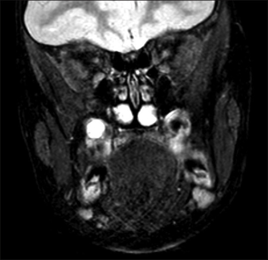
Coronal T2 FATSAT image showing accessory parotid gland tissue (marked with arrow) bilaterally overlying masseter muscle
Figure 3.
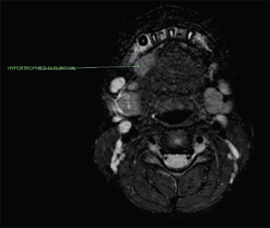
Axial T2 FATSAT image showing well-defined soft tissue in the right sublingual region isointense to salivary gland tissue representing hypertrophied sublingual gland
A non-contrast computed tomography (CT) scan was also performed to look at bony orofacial structures and it confirmed the MRI findings of the salivary glands. Associated gray matter heterotopia was seen in the left frontal region. The note was also made of cavum septum pellucidum [Figure 4]. Supernumerary teeth were seen along the alveolar border of the maxilla with a solitary mandibular central incisor [Figure 5].
Figure 4.
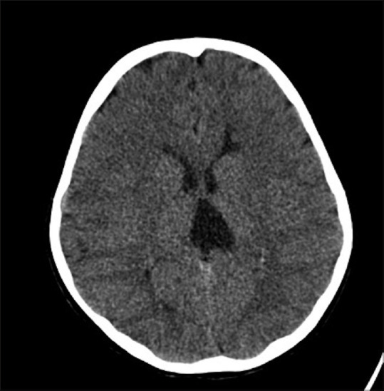
Axial plain CT scan showing a small subependymal heterotopia in the left frontal lobe. There is associated cavum velum interpositum
Figure 5.
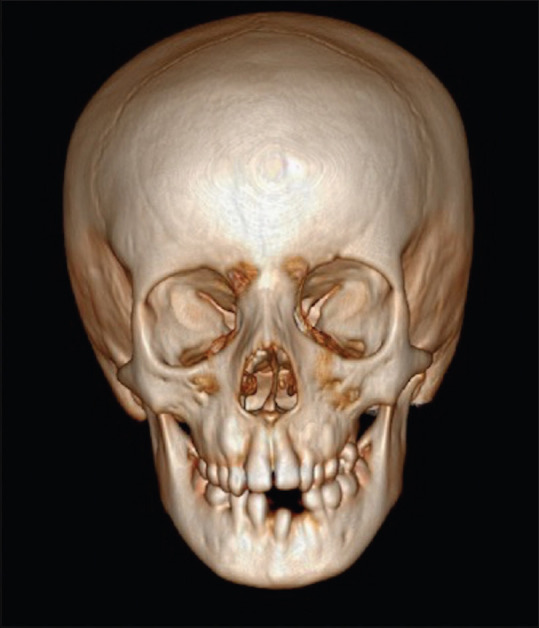
3D reconstructed image showing central solitary incisor
Diagnosis
Aplasia of unilateral parotid with hypoplasia of contralateral parotid with hypertrophy of unilateral sublingual gland.
Discussion
Unilateral or bilateral agenesis of the parotid gland is an uncommon condition with unclear aetiology. Bilateral agenesis is relatively more common and is associated with other developmental craniofacial anomalies.[1]
Gruber reported the first case of salivary gland agenesis.[2] As the condition is asymptomatic by itself, the exact incidence is difficult to ascertain. Other major and minor salivary glands being the main source of saliva, parotid gland agenesis does not result in xerostomia.
Only 22 cases of unilateral salivary agenesis have been reported excluding the present case. Teymoortash and Hoch reported a case of unilateral agenesis and analysed 21 available cases from the literature. They noted that the condition showed no sex predilection with age at the time of diagnosis ranging from 50 to 75 years. Their analyses found the unilateral absence of the gland to be twice as common on the left, in contrast to our case where it was on the right.[1]
In most of these cases, the authors reported a facial asymmetry or a swelling of the contralateral parotid gland which was believed to be due to compensatory functional hypertrophy.[1] The present case is unique in this regard as it showed the contralateral parotid to be hypoplastic with the hypertrophied sublingual gland. Four out of the 22 cases analysed were noted to have ipsilateral accessory parotid tissue. Our case showed the presence of accessory parotid tissue bilaterally. No associated with heterotopia in the brain and cavum septum pellucidum has been reported in the literature so far.
The congenital cleft tongue is also a rare disorder which is usually seen with syndromes such as orofacial digital syndrome, facial clefts, and also associated with abnormal tissue growths, usually hamartomas.[3] This is also seen in association with supernumerary teeth in oro-facial dental syndromes, as seen in our case, but none of the other features of the syndromes were noted.
We were unable to classify our findings into any well-defined syndrome. The patient does not fit into any of the oro-facial-dental syndromes with a combination of anomalies not reported in the literature so far to the best of our knowledge.
Declaration of patient consent
The authors certify that they have obtained all appropriate patient consent forms. In the form the patient(s) has/have given his/her/their consent for his/her/their images and other clinical information to be reported in the journal. The patients understand that their names and initials will not be published and due efforts will be made to conceal their identity, but anonymity cannot be guaranteed.
Conclusion
Agenesis of the parotid gland is an uncommon congenital anomaly usually associated with different craniofacial anomalies. It is essential to evaluate orocraniofacial structures in all individuals with parotid agenesis.
Financial support and sponsorship
Nil.
Conflicts of interest
There are no conflicts of interest.
References
- 1.Teymoortash A, Hoch S. Congenital unilateral agenesis of the parotid gland: A case report and review of the literature. Case Rep Dent 2016. 2016 doi: 10.1155/2016/2672496. 2672496. [DOI] [PMC free article] [PubMed] [Google Scholar]
- 2.Gruber W. Congenitaler mangel beider glandulae submaxillares bei einem wohlgebildeten, erwachsenen Subjecte. Arch für Pathol Anat und Physiol und für Klin Med. 1885;102:9–11. [Google Scholar]
- 3.Nutalapati R, Jayasuriya N. Salivary hamartoma with a bifid tongue in an adult patient. Natl J Maxillofac Surg. 2018;9:61–63. doi: 10.4103/njms.NJMS_28_17. [DOI] [PMC free article] [PubMed] [Google Scholar]