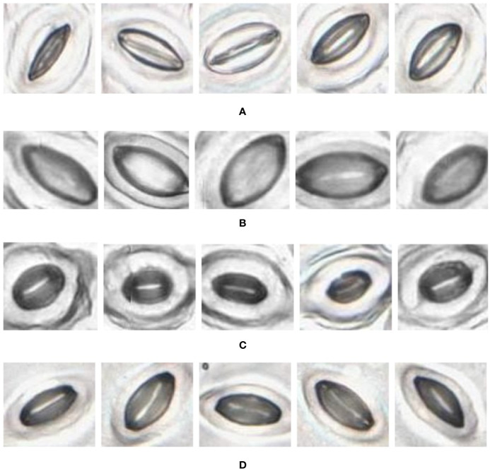
Figure 10.
Individual stomata captured from traditional samples using the slide scanner. Guard cell boundaries and background epidermal cells are clearly visible. (A) Vitis vinifera L. x V. rupestris Scheele ‘Ganzin Glory’. (B) Prunus armeniaca ‘Moorpark’. (C) Citrus sinensis L. Osbeck ‘Valencia’. (D) Vinca major L. ‘Periwinkle’.

