Figure 3.

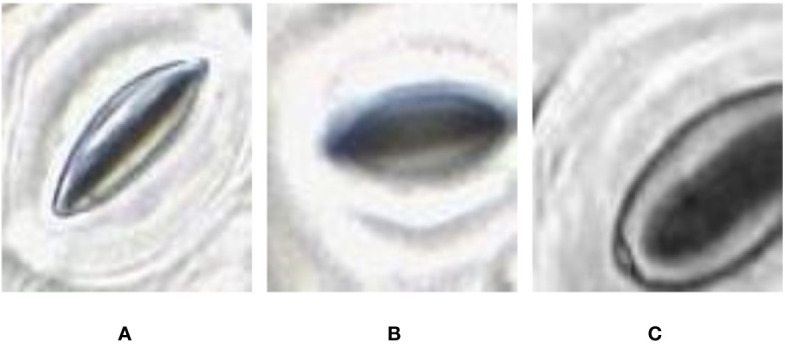
Quality variation of the stomata captured at different parts of the microscope image. (A) sharp image. (B) blurry image (C) partially captured image.

Quality variation of the stomata captured at different parts of the microscope image. (A) sharp image. (B) blurry image (C) partially captured image.