Figure 1.
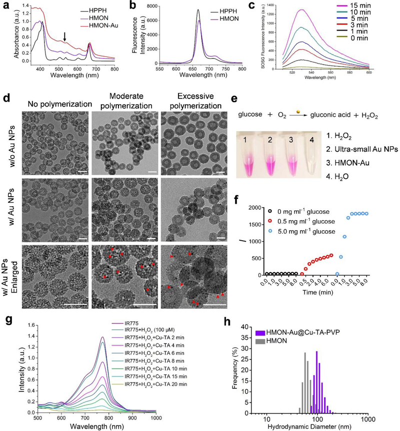
Preparation and characterization of HPPH hybridized HMONs (abbreviated as “HMON”), HMONs immobilized with the ultra-small Au NPs (HMON-Au) and HMON-Au@Cu-TA-PVP nanoparticles. (a) UV-Vis absorption of HPPH, HMON and HMON-Au. (b) Fluorescence spectra of HPPH and HMON. (c) Fluorescence spectra of the SOSG solution incubated with HMON over increased irradiation time. (d) Morphologies of HMON with no polymerization, HMON with moderate polymerization, and HMON with excessive polymerization (the upper panel, w/o Au NPs) detected by TEM; morphologies of HMON with no polymerization, HMON with moderate polymerization, and HMON with excessive polymerization incubated with the ultra-small Au NPs (the middle and lower panel, w/ Au NPs) detected by TEM. Scale bar, 50 nm. (e) The digital images of H2O2 production after indicated treatment detected by Hydrogen Peroxide Assay Kit. (f) The quantification of H2O2 production in the presence of HMON-Au at indicated concentration of glucose. (g) UV-Vis absorption of IR775, a radical indicator, after indicated treatment. (h) Size distribution of HMON and HMON-Au@Cu-TA-PVP nanoparticles measured by DLS.

