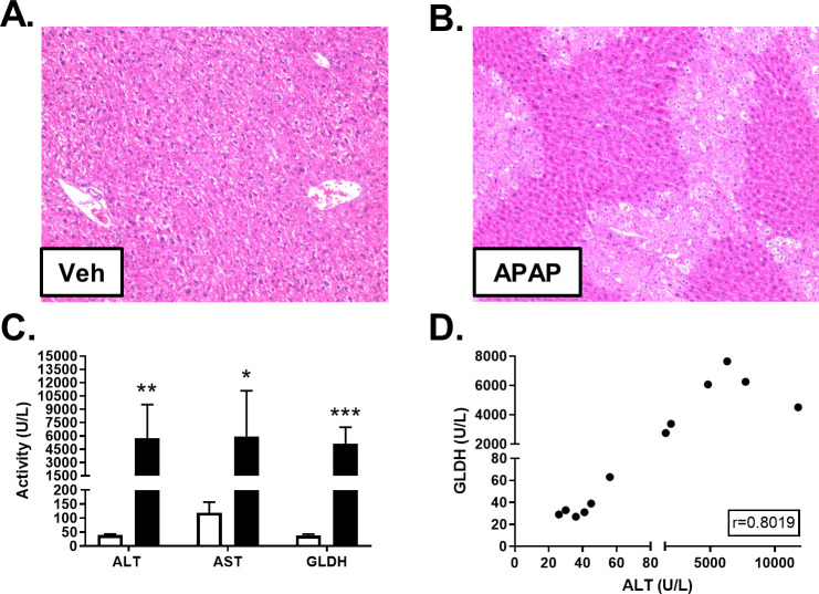
Fig 2. Effect of APAP on liver histology and biomarkers.
Male C57BL/6J mice received a single intraperitoneal administration of vehicle (Veh; 0.9% saline) or APAP (300 mg/kg). Liver tissue and blood were collected 24h post-dosing. H&E staining revealed APAP-induced hepatocellular necrosis. Representative photomicrographs at 10X magnification are shown for vehicle- (A) and APAP-dosed mice (B). ALT, AST and GLDH were quantified in freeze/thawed plasma samples from Veh (white) and APAP-treated (black) mice. Significance was *p<0.05, **p<0.01, or ***p<0.001 (C). Pearson’s r was calculated to explore the correlation between ALT and GLDH quantities measured in plasma (D).

