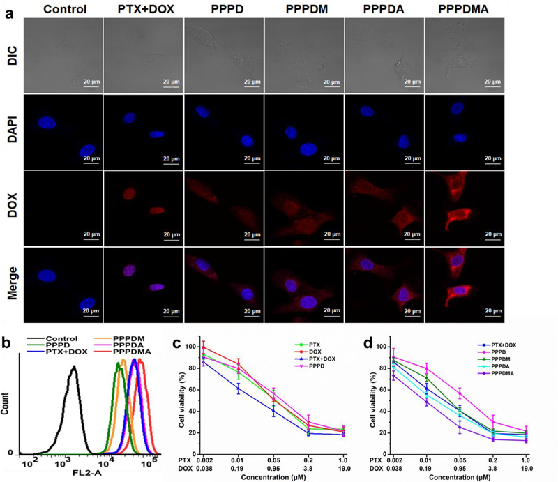
Figure 2.
The in vitro targeting effect of the PPPDMA nanogel. (a) Representative CLSM images of SKOV-3 cells after various treatments for 3h. Cell nuclei were stained with Hoechst 33342. The red fluorescence is from DOX, and the blue fluorescence is from Hoechst 33342. The scale bars are 20 μm. (b) Flow cytometry analysis of SKOV-3 cells after various treatments for 3h. (c) Cell viability of SKOV-3 cells after various treatments with free PTX, free DOX, a combination of PTX and DOX, and PPPD nanogels for 72 h. (d) Cell viability of SKOV-3 cells after various treatments with the combination of free PTX and DOX, PPPD, PPPDM, PPPDA, and PPPDMA nanogels for 72 h. Data represent the means ± SD, n=4.

