Figure 3.
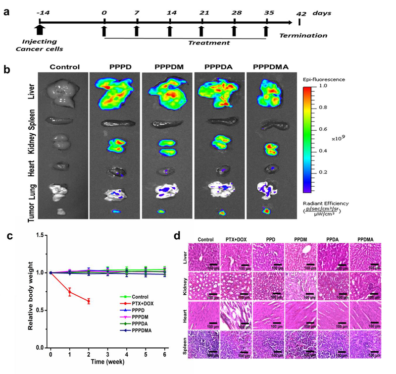
Treatment schedule, biodistribution, and systemic toxicity of the nanogels in a peritoneal metastatic ovarian tumor mouse model. (a) Schedule of treatment of the nanogels. (b) Ex vivo fluorescence images of mice organs and tumors after treatments with various nanogels. (c) Body weight changes over the duration of treatment. Mice in the PTX and DOX group died after receiving 2 weeks of treatment. (d) Representative histological features of the tissues after 6 weeks of nanogel treatment. Mice in the PTX and DOX group only received 2 weeks of treatment.

