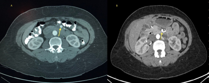
Figure 2.
(Left) Axial view of computed tomography angiogram showing a cross-section of a long-segment intimal dissection flap (arrow) in the distal superior mesenteric artery (SMA). (Right) Follow-up computed tomography angiogram with axial image showing improvement of the distal SMA dissection (arrow)

