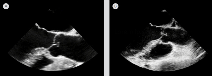
Figure 2.

Transesophageal echocardiogram (TEE). (A) TEE in the long axis view performed on admission with no vegetations on the aortic valve. (B) Repeat TEE performed on admission day 5 indicating a new mobile vegetation on the right coronary cusp of the aortic valve, measuring 0.5 cm by 0.4 cm, even though the patient was on antimicrobial therapy and heparin.
