Figure 1.

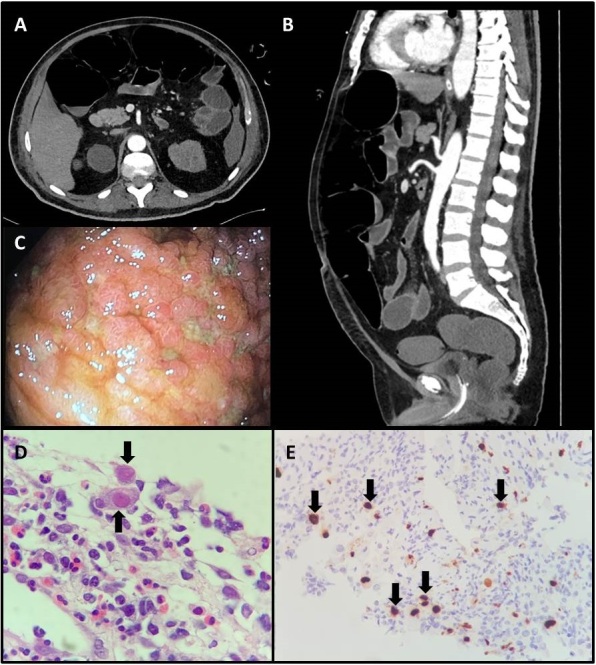
(A,B) Computed tomography scan of the abdomen revealed jejunal thickening with no evidence of mesenteric ischaemia. (C) Colonoscopy findings showed an extensive ulcerated area, measuring about 5.0 cm, affecting 80% of the ileum circumference. (D) Biopsy of the ileum revealing extensive ulceration with fibrino-leucocytic exudate and endothelial cells with viral cytopathic changes (black arrows, ×400). (E) Immunohistochemistry for cytomegalovirus showed positive staining in several cells (black arrows, ×100)
