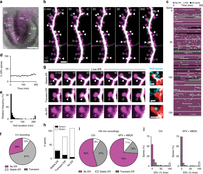
Fig. 1. ER dynamics is regulated by synaptic activity.
a Organotypic hippocampal slice transfected using a Helios gene gun. Overlay of bright field and fluorescence images shows expression of tdimer2 (magenta) and ER-EGFP (green fluorescence, white in overlay) in a few CA1 neurons. Scale bar: 500 μm. Experiment was reproduced in ten slices. b Two-photon maximum intensity projections of a dendritic branch from a hippocampal CA1 pyramidal neuron transfected with tdimer2 (magenta) and ER-EGFP (green, white when over magenta) followed at 10 min intervals up to 5 h. Filled and empty arrowheads denote presence and absence of ER in a few representative spines, respectively. Scale bar: 2 μm. Horizontal structure is an axon. Experiment was reproduced on three slices with similar results. c Score sheet of dendritic spines monitored over 5 h (n = 153 spines, 2 neurons, 2 slices). Magenta: spine without ER; white: spine with ER; black: spine head not visible/not analyzed. d Percentage of spines from c containing ER over time. e Histogram of ER visit duration over a 5 h period (from c). f Percentage of spines with stable ER, receiving at least one visit (transient ER) or no visit (no ER) within 5 h of observation (from c). g Three examples (two-photon time series, maximum intensity projections) of spines with stable (top), transient (middle), or no ER (bottom), followed by correlative confocal images (maximum intensity projections) of the same spines (native tdimer2 fluorescence, red) after fixation of the tissue and immunostaining against synaptopodin (cyan). Scale bars: 1 µm. Note synaptopodin clusters inside (white) as well as outside (cyan) the transfected neuron (red), as the antibody labels synaptopodin in the entire neuropil. Experiment was reproduced on two slices. h Synaptopodin clusters were detected in 90% of stable ER+ spines, in 16% of transient ER spines, and in 4% of spines with no ER visit (n = 220 correlatively imaged spines, 2 cells, 2 slices). i Over a period of 100 min, 45% of spines were visited by ER at least once. Blocking excitatory transmission (APV + NBQX) reduced the fraction of transiently visited spines from 37% (n = 262 spines, 3 neurons, 3 slices) to 15% (n = 192 spines, 2 neurons, 2 slices). j Residence time of ER in spines, under control conditions (n = 262 spines, 3 neurons, 3 slices) and with blocked excitatory transmission (n = 192 spines, 2 neurons, 2 slices) (p = 0.0005, two-sided Mann–Whitney test). Source data are provided as a Source Data file.

