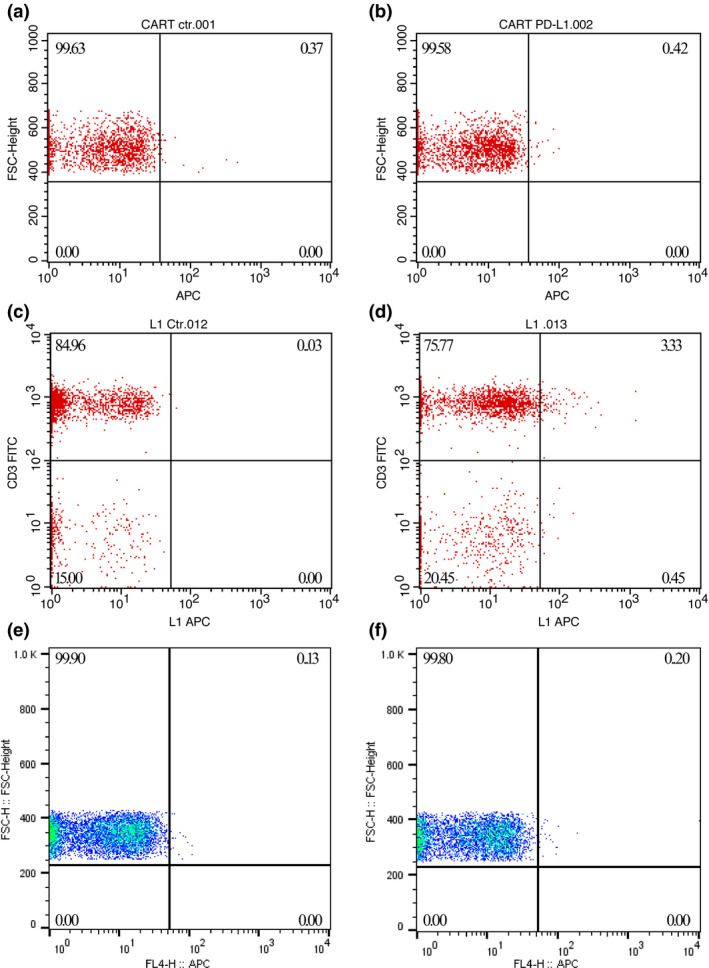
Figure 5.

PD‐L1 CAR‐T cells proliferated greatly after transfusion in the patient’s circulation.(upper panels) No PD‐L1 CAR‐T cells were detected on Day +11, when the patient was discharged from the hospital after CAR‐T cell infusions. (a) Patient’s PBMCs stained with APC‐labelled streptavidin alone. (b) Patient’s PBMCs stained with both biotinylated PD‐L1::human Fc fusion protein and APC‐labelled streptavidin. (middle panels) Approximately 3.30% of the total T cells were CAR positive on Day +29. (c) Patient’s PBMCs stained with FITC CD3 and APC‐labelled streptavidin. (d) Patient’s PBMCs stained with FITC CD3 and both biotinylated PD‐L1::human Fc fusion protein and APC‐labelled streptavidin. (lower panels) PD‐L1 CAR‐T cells were undetectable after the patient developed ALI/ARDS on Day +48. (e, f) The staining patterns were the same as those in a and b, respectively.
