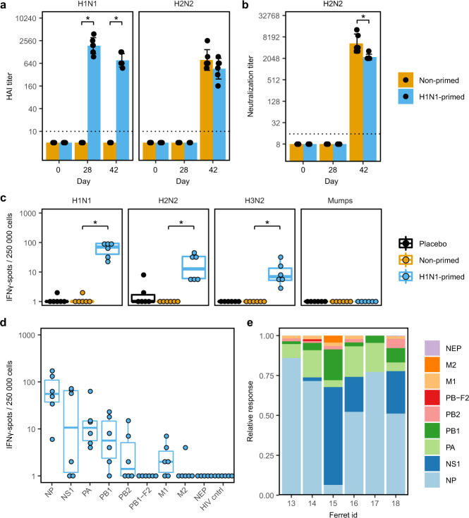
Fig. 3. H1N1-priming induced H2N2-reactive T cells but no H2N2-neutralizing antibodies.
a, b Sera from primed and non-primed ferrets was tested for antibody responses against A/California/07/2009 (H1N1) and A/Singapore/1/57 (H2N2) before H1N1-priming (day 0), before H2N2 infection (day 28) and 2 weeks after H2N2 infection (day 42). Bars depict mean ± SD. a Hemagglutination-inhibition (HAI) titers against H1N1 and H2N2. b Virus neutralization titer against H2N2. c–e IFNγ-responses by ELISpot after ex vivo stimulation of PBMCs obtained 28 days after H1N1-priming. Responses are corrected for background signals (minus medium stimulation). c Responses against H1N1, H2N2, A/Switzerland/97/2013 (H3N2), and mumps virus. d Responses of H1N1-primed animals to peptide pools spanning the internal proteins of H2N2 and the control HIV gag protein. Non-primed and placebo animals were negative for all peptide pools tested but were left out for visualization purposes. e For each ferret shown in d, relative responses against the different influenza proteins was calculated. Boxplots depict the 25, 50, and 75% quantile where the upper and lower whiskers extend to the smallest and largest value respectively within 1.5* the inter quartile ranges. In all plots n = 6. * indicates significant differences (p < 0.05) between non-primed and H1N1-primed groups after correction for multiple testing.

