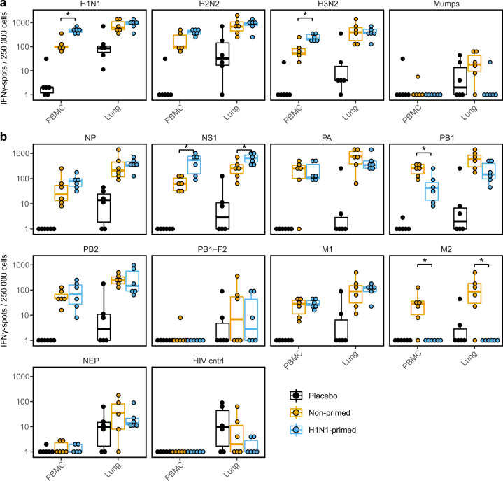
Fig. 6. Primed animals show increased responses to influenza A virus.
a, b Lymphocytes isolated from blood or lung were stimulated with multiple influenza virus subtypes (a) or H2N2 peptide pools (b) in an ELISpot assay for 20 h. Responses are corrected for background signals (minus medium stimulation). In all panels, n = 5–6. Boxplots depict the 25, 50, and 75% quantile where the upper and lower whiskers extend to the smallest and largest value respectively within 1.5* the inter quartile ranges. Asterisk indicates significant differences (p < 0.05) between non-primed and H1N1-primed groups after correction for multiple testing.

