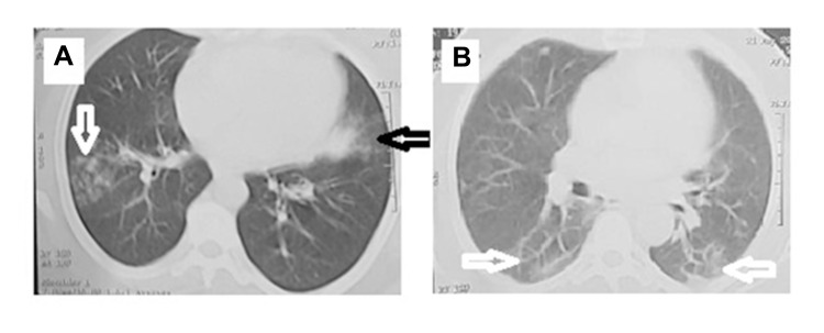
Figure 2.
Radiological examination of COVID-19 patient. (A) COVID-19 pneumonia in a 48-year-old female who complained of intermittent fever, shortness of breath and headache for 3 days before admission. Plain computed tomographic scan of the lung shows an area of ground-glass attenuation on one side (black arrow) and tree in bud appearance on the other side(white arrow). (B) COVID-19 pneumonia in a 57-year-old male who complained of, headache, vomiting, diarrhea for 5 days before admission. Plain computed tomographic scan of the lung that shows an area of crazy paving with interlobular septal thickening in both lower lobes (white arrows).

