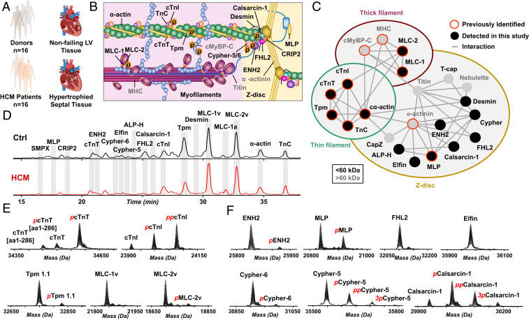
Fig. 1.
A complex sarcomeric proteoform landscape. (A) Control tissue from left ventricle (LV) of nonfailing donor hearts (Ctrl, n = 16) and HCM tissues procurred via surgical septal myectomy procedure (HCM, n = 16). (B) Schematic representation of cardiac sarcomere, consisting of thin (green) and thick filaments (pink) flanked by Z-disk. (C) Sarcomeric protein interactome showing a complex network of interactions between myofilament and Z-disk proteins. (D) Representative base peak chromatograms showing separation and detection of major sarcomeric proteins by LC-MS for Ctrl and HCM tissues. (E and F) Representative deconvoluted mass spectra showing the proteoforms of (E) myofilament and (F) Z-disk proteins.

