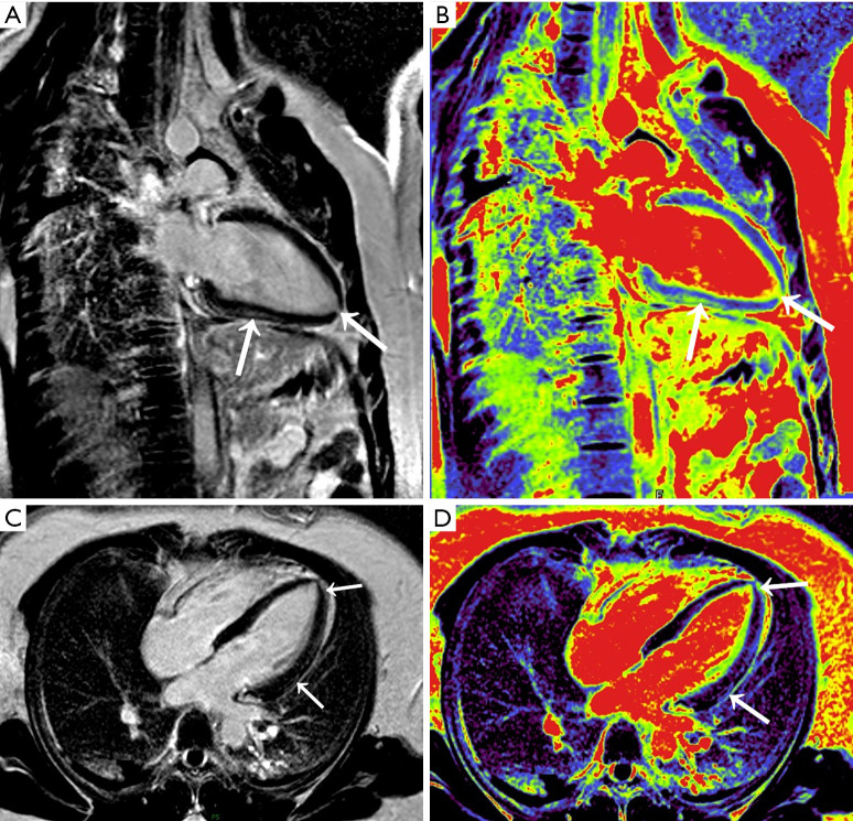
Figure 2.
Original two-chamber (A), pseudo-color two-chamber (B), original four-chamber (C), and pseudo-color four-chamber (D) delayed-enhancement T1-weighted multishot gradient-echo IR magnetic resonance images of myocarditis in a 40-year-old woman. The band-like subepicardial LGE (arrows) of the left-ventricular lateral and inferior walls associated with nodular predominating in the apical segment can be seen. LGE, late gadolinium enhancement.

