
Figure 2. Pathologic tumor response correlates with tumor PD-L1 and immune infiltrates.
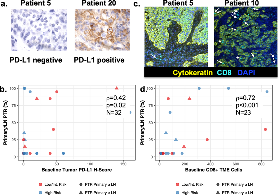
(A) Representative PD-L1 staining of tumor biopsies at baseline. (B) PD-L1 H-score correlated with pathologic tumor response (PTR). Baseline PD-L1 primary tumor expression levels by IHC and percent PTR were significantly positively correlated for 32 evaluated patients (rho=0.43; 95%CI 0.079–0.668) (C) representative multiplex immunofluorescence (MIF) images showing patient 5 with minimal CD8+ T cell infiltrates and Patient 10 with higher CD8+ T cell infiltrates (white arrows) in baseline biopsies. (D) Extent of PTR was correlated with number of CD8+ T cells in the baseline biopsy tumor microenvironment (TME). Baseline number of TME CD8+ T cells assessed by MIF and percent PTR were significantly positively correlated for 23 evaluated patients (rho=0.72; 95%CI: 0.443–0.875).