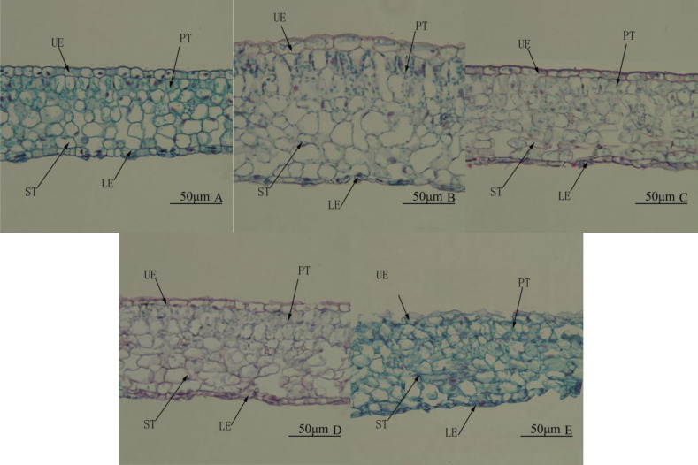
Figure 4. Leaf anatomy. Cross-section of the middle part of the leaf blade of Camellia oleifera cultured under light of different spectral quality, photographs were taken at 20 × magnification.
Photographs were taken at 20 × magnification. (A) White light (WL); (B) red:blue (R4:B1); (C) blue light (BL); (D) red:blue (R1:B4); (E) red light (RL). UE, upper epidermis; LE, lower epidermis; PT, palisade mesophyll tissue; ST, spongy mesophyll tissue. Scale bar = 50 µm.

