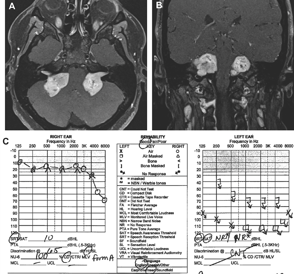
Figure 1. Magnetic Resonance Imaging (MRI) of Bilateral Vestibular Schwannoma (VS).
Axial [A] and coronal [B] cuts of a T1-weighted contrast-enhanced MRI demonstrate bilateral VS in a 25 year-old female with Neurofibromatosis Type 2. [C] The right VS is significantly larger than the left VS, however the hearing is overall preserved on the side with the larger tumor.

