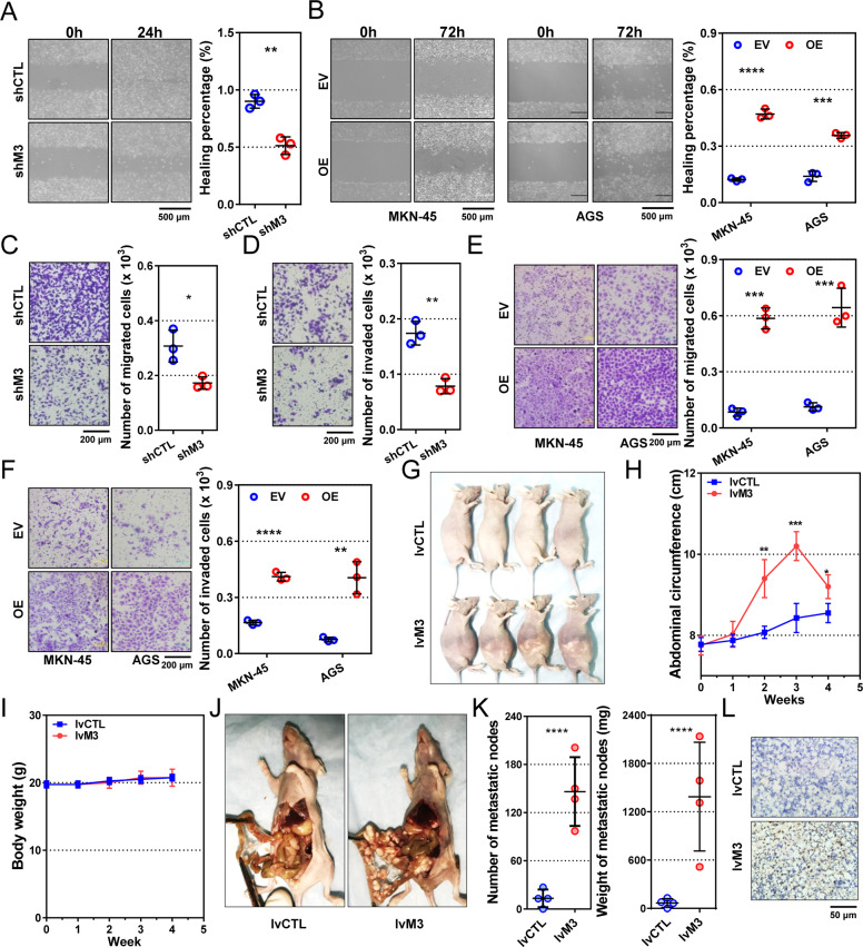
Fig. 3. METTL3 promotes gastric cancer cell motility and peritoneal colonization.
a, b Wound-healing assays of METTL3-reducing (shM3) HGC-27, METTL3-overexpressing (OE) MKN-45, and AGS, and their corresponding control (shCTL and EV) cells. c, d Migration and invasion assays of METTL3-reducing HGC-27 cells. e, f Migration and invasion assays of METTL3-overexpressing MKN-45 and AGS cells. Representative images on the left, and quantification bars on the right in a–f (n = 3 for each group). g Mice with peritoneal implant nodes derived from METTL3-overexpressing (lvM3; n = 4) and control (lvCTL; n = 4) MKN-45 cells. h, i Curves of abdominal circumferences and body weights of the peritoneal metastasis models. j Representative pictures of the peritoneal implants from stable METTL3-overexpressing and control MKN-45 cells. k Number (left) and weight (right) of implanted nodes in peritonea of mouse models. Each dot represents one sample. l Representative pictures of METTL3 expression in peritoneal nodes by IHC. Data are presented as mean ± SD. *P < 0.05; **P < 0.01; ***P < 0.001; ****P < 0.0001.

