
Figure 5.
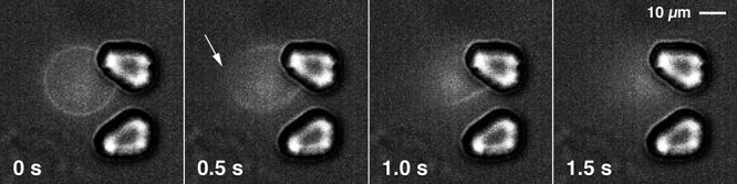
Snapshots of a video sequence of a giant POPC vesicle (diameter of 20 μm) with membrane-incorporated DiD dye, floating in the microfluidic chip chamber in the presence of 50 μM peptide 6, the most active peptide on S. aureus SH1000. The flow was from left to right at a rate of 0.5 μL min–1. Two micropillars acted as a hydrodynamic trap for the vesicle. The white arrow indicates the position at which the lipid membrane of the vesicle started to rupture upon exposure to peptide 6.