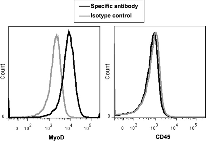
Fig. 2.
The immunophenotype of myoblast expanded from skeletal muscles of donors. To evaluate the expression of MyoD and CD45 on myoblast (passage 1), fluorescent-labeled antibodies against MyoD and CD45 were used to stain the myoblast, and then the cell staining was analyzed by flow cytometry. Representative expression of MyoD and CD45 on myoblast expanded from one donor

