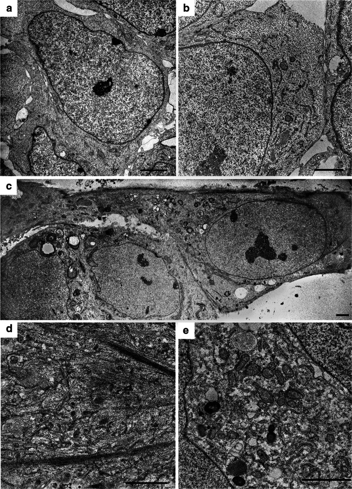
Fig. 4.
Ultrastructural transformation of the cells during differentiation. a An overview of a pluripotent cell containing a large nucleus surrounded by a narrow layer of cytoplasm. b Part of a pluripotent cell at higher magnification showing the following: mitochondria, short ER cisternae and cytoplasm with organelles. c An overview of cells on the 5th day of differentiation: triangular cells were observed with an emerging branch and cytoplasm filled with organelles (on the right). d A branch fragment of the cell after a first stage of differentiation (12–13 days) with free-lying and bundled neurofilaments. e A part of the cytoplasm on day 12–13 with a high density of mitochondria, ER cisterns, and autolysosomes. The scale bar is 2 µm

