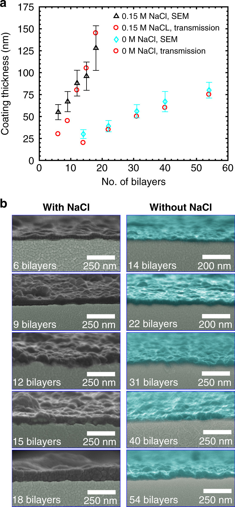
Fig. 2. Comparison of the capillary polymer coatings obtained by the PEs dissolved in deionized water and in buffer containing 0.15 M NaCl.

a Polymer coating thickness versus the number of PE bilayers. The coating thickness is evaluated from SEM images (SEM) and obtained by fitting the shifts in the fibre transmission spectra through the condition of Fabry–Perot resonances (transmission) (see the Supplementary information). Polymer coatings are included in the theoretical model as additional concentric layers of equal thickness on the core capillary. The thickness is fitted to obtain the coincidence of the minima positions for both the experimental and calculated transmission spectra. Error bars show the coating roughness. b SEM micrographs of the core capillaries functionalized with different numbers of PE bilayers. Left column: PEs in deionized water with 0.15 M NaCl. Right column: PEs in pure deionized water. Pseudocolour indicates the polymer coating
