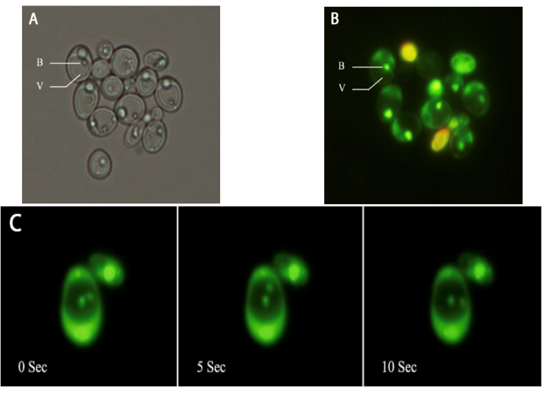
Fig.1.
Light and fluorescence microscopy of yeast. a) Light microscopy of yeast cells shows intracellular bacteria (B) inside yeast’s vacuole (V). b) Live bacteria (B) appeared as green spots in the vacuole (V) of stained yeast cells. c) Photographs taken at three-time intervals (0, 5, and 10 seconds) show the moving bacteria. Original magnification x 1000.

