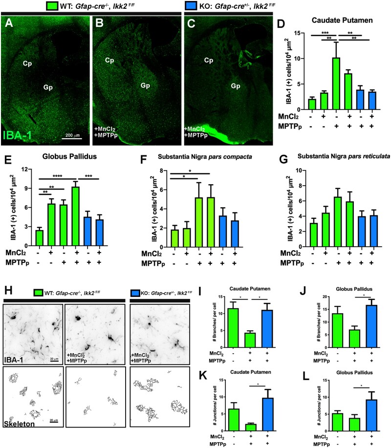
Figure 5.
Astrocyte-specific deletion of I kappa B kinase 2 (IKK2) suppresses a reactive phenotype in microglia following sequential exposure to MnCl2 and 1-methyl-4-phenyl-1,2,3,6-tetrahydropyridine (MPTP). The number of microglia in mice sequentially exposed to MnCl2 during juvenile development and to 1-methyl-4-phenyl-1,2,3,6-tetrahydropyridine and probenecid (MPTPp) as adults was analyzed by automated quantitation of tissue sections immunolabeled with anti-IBA1 (green), as depicted in montage images of (A) (wildtype) WT—control, (B) WT—MnCl2/MPTPp, and (C) (knockdown) KO—MnCl2/MPTPp treatment groups. The total number of IBA1+ cells/104 µm2 was quantified in the caudate-putamen (Cp) (D), globus pallidus (Gp) (E), substantia nigra pars compacta (F) and substantia nigra pars reticulata (G) for all experimental groups (*p < .05; **p < .01, ***p < .001, ****p < .0001; N = 5–6 animals/group). H, Morphological changes in microglia between treatment groups were analyzed by image skeletonization of IBA1+ cells using Image J, as depicted in representative images of WT—Control, WT—MnCl2/MPTPp, and KO—WT + MnCl2/MPTPp. Parameters quantified in each experimental group included the number of branches/cell in Cp/Gp (I, J), and the number of junctions/cell in Cp/Gp (K, L) (*p < .05; N = 7 animals/group).

